Batavialab - PT. Mitra Batavia Semesta
| (+62 21) 8371 7846 | (+62 21) 2868 1579 | +62 877 3917 7737 | sales1@batavialab.com |
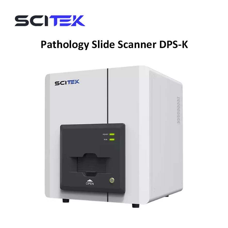
Pathology Slide Scanner DPS-K Scitek
Pathology Slide Scanner Advantage:
1. Segmented Power Control for Safe Operation. High and low voltage circuits are independently controlled during startup to prevent "surge" damage to sensitive electronic modules and ensure operator safety.
Magnetic Slide Holder. Magnetic design ensures precise positioning and smooth loading/unloading, preventing slide damage or slippage.
2. High-Speed, High-Precision X/Y/Z Stage System. Imported magnetic levitation linear motor: Speed up to 3.2m/s, acceleration up to 8g. Full closed-loop control system: X/Y-axis resolution 100nm, Z-axis resolution 50nm.
3. Advanced Scanning Software Features. Instant preview: View scanned images immediately for diagnosis. Real-time positioning: Microscope-like navigation to locate and display scanned regions.
4. Dynamic parameter display: Shows real-time positional and tissue information. Multi-tissue recognition: Intelligently segments and scans multiple tissue blocks on a single slide, merging them into a unified digital image.
5. Viewing Software Capabilities. Slide management: Automatically detects, sorts, searches, and categorizes slide files. Color-coded navigation: Tracks viewing paths at different magnifications. ROI annotation: Marks regions of interest with customizable shapes, colors, and line widths; exports annotations to Excel. High-resolution capture: Supports 2300dpi screenshots (partial or full-screen) for publication; allows labeling and exporting.
-----------------------------------------------------------------------------------------------------------
|
Model |
DPS-K |
|
Loading Capacity |
5 slides |
|
Slide Size |
26mm×76mm |
|
Objective Lens |
Plan Apochromatic (APO) 20× objective, Numerical Aperture NA0.75 |
|
Core Imaging Camera |
5-megapixel CMOS area-array camera, 2/3 inch |
|
Scan Magnification |
20×/40× automatic switching |
|
Scan Method |
Area-array scanning |
|
Scan Area |
≥24mm× 50mm |
|
Scan Speed |
20×: <30 seconds |
|
Focusing Method |
Auto-focusing and manual fine-tuning |
|
Specimen Identification |
Supports standard barcodes, QR codes, etc. |
|
Tissue Recognition |
Automatically detects tissue shape and region; skips blank areas; intelligently segments and scans multiple tissue blocks on a single slide |
|
Multi-layer Fusion Scan |
Stores individual layer images for cellular morphology observation; fuses clearest regions from each layer into a composite image |
|
Scan Output |
24-bit color, real-time preview |
|
Data Storage |
Local and cloud storage |
|
Shipping Size(W×D×H)(mm) |
860×850×1450 |
|
Weight |
153kg |
Produk
Lokasi Kami
Batavialab - PT. Mitra Batavia Semesta
| PT. Mitra Batavia Semesta Banjaran Pucung No. 171 RT.002 RW.005 Kelurahan Cilangkap, Kecamatan Tapos Kota Depok, Jawa Barat |
|
| 021 8371 7846 / 021 2868 1579 | |
| 087739177737 | |
| sales1@batavialab.com |