
Batavialab - PT. Mitra Batavia Semesta
| (+62 21) 8371 7846 | (+62 21) 2868 1579 | +62 877 3917 7737 | sales1@batavialab.com |
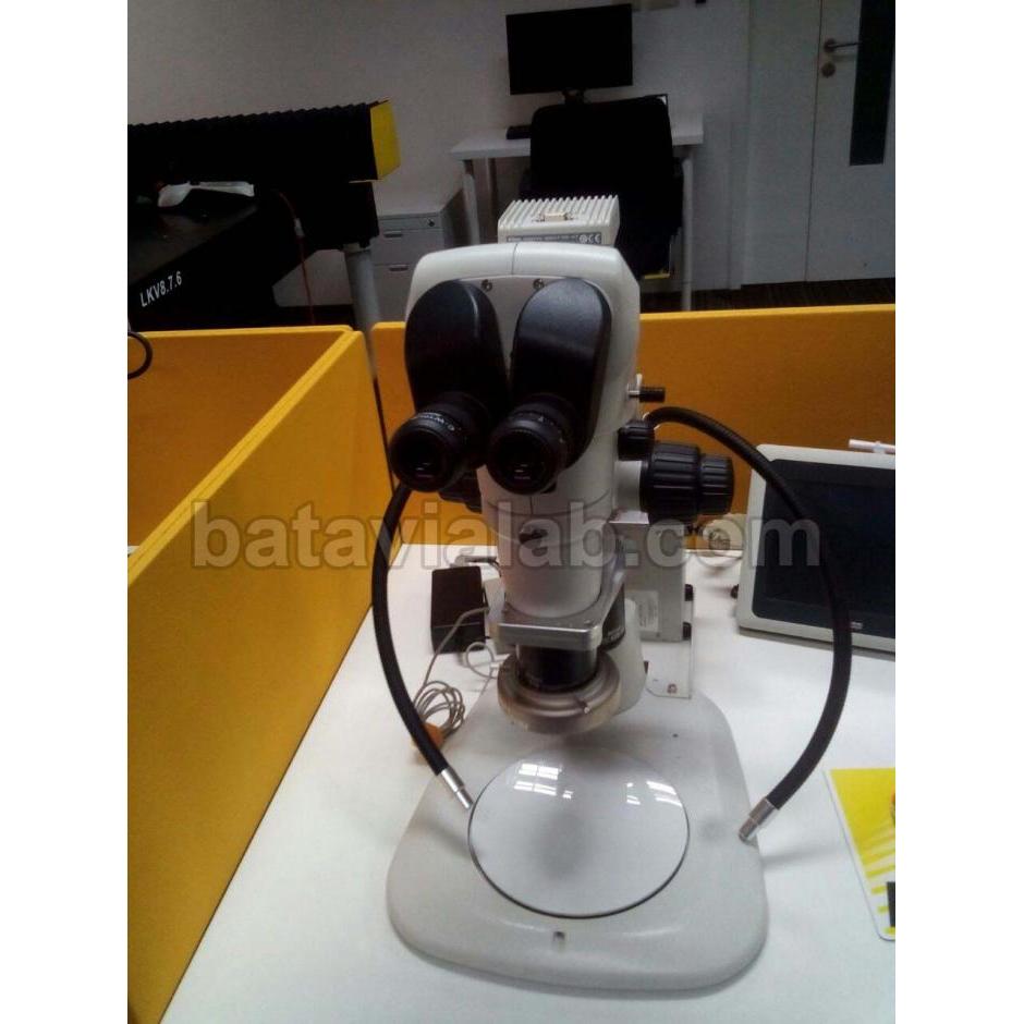
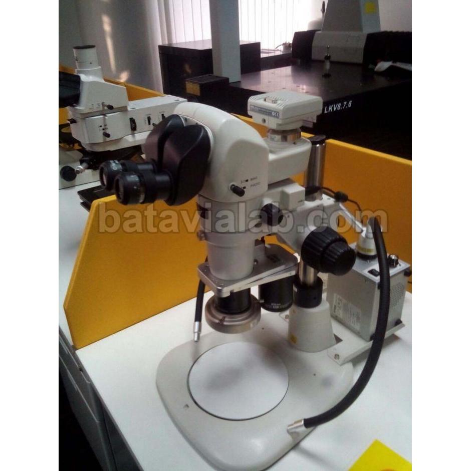
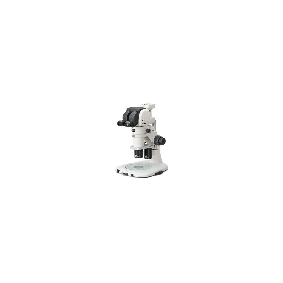
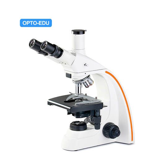
Mikroskop Stereo SMZ1270i
An advanced stereomicroscope featuring an expanded zoom range, remarkable image sharpness, and intelligent image acquisition features.
To meet and exceed the needs to observe minute structures while covering a wide field of view in the biological fields where biological body screening, injection and observation are conducted every day, the SMZ1270i was developed to feature expanded zoom ratio, high operability, intelligent image acquisition features and excellent optical performance.
Key Features
Best-In-Class Zoom Ratio

With a best-in-class zoom ratio of 12.7x (0.63x-8x), the SMZ1270i covers an even greater magnification range than previous models. At the lowest magnification, the actual field of view will go up to 35mm*, allowing easy observation of an entire 35mm petri dish.
* when 1x objective and 10x eyepieces are used (excluding Coaxial EPI illumination).

0.63x zoom

8x zoom
High Quality Images
Amazingly clear and bright images with reduced chromatic aberrations can be acquired throughout the extended zoom range through the newly developed “Plan Apo WF” and “ED plan WF” objectives.


Apochromat optics

Conventional optics
New Objectives Optimized for Widefield Observation at Low Magnification

In combination with the newly developed WF series objectives, the SMZ1270i offers a wide and uniformly bright viewfield even at low magnifications. In addition, a 0.75x objective is now available, expanding the lineup of low magnification objectives.
Versatile Double Nosepiece
The double nosepiece allows for seamless observation in a wide range of magnifications. In addition, changing objectives has never been easier.
By changing the nosepiece from stereo-position (stereo view) to
mono-position (on-axis view), you can easily acquire images suited for
measuring and processing.

On-axis view

P-RN2 Nosepiece
Improved Ergonomics

There are several eyepiece tubes to fit various configurations, from a simple setup to a system with some intermediate modules or digital cameras. With an ergonomic eyepiece tube, the observation angle can be adjusted from 0-30 degrees, allowing for comfortable observation postures even if combined with intermediate modules like illuminators or teaching heads. Furthermore, the Plain Stands and Diascopic LED Stands adapted a slim design for easier operation and exchange of specimens and samples.
Advanced Digital Imaging Capabilities

Intelligent Nosepiece P-RN2
In combination with the DS-L3 Camera Control Unit, NIS-Elements imaging software, and P-RN2 Intelligent Nosepiece, image acquisition is now more efficient, with the objective magnification and zoom ratio information displayed on the monitor.
 Stand-alone Control Unit DS-L3 |
 PC-use Control Unit DS-U3 (with NIS-Elements) |
 |
 |
Expandable with a Wide Range of Accessories
To support a variety of observation methods and applications used for research and inspection, an assortment of accessories, such as illumination units, stands, and eyepiece tubes, are available. By incorporating a “Fly Eye Lens” with the Epi-Fluorescence illuminator, uniform brightness over the entire field of view is realized, which helps in observing specimen that needs a wide magnification range. Nikon also offers several digital cameras ideally suited different stereo microscope applications.
Diascopic illumination stands have been renewed. The slim LED base design provides easy access to specimen and easy operation. As with the SMZ25 and SMZ18, the diascopic illumination base features Nikon’s very own oblique illumination, the OCC illuminator. The built-in feature offers high contrast observation for transparent samples such as oocytes used in the IVF application.
| OCC Illumination | Diascopic brightfield illumination | |
 |
 |
 With the LED Diascopic Illumination Base and P-DSF32 Fiber Diascopic Illumination Stand, image contrast under OCC illumination can be easily adjusted. |
| OCC illumination boosts the contrast of transparent sample structures. Hemicentrotus pulcherrimus in two-cell stage | ||
 Fibers |
 Semiconductor |
 Brain of adult drosophila excited with GFP Image courtesy of Hokto Kazama, Ph.D., Circuit Mechanisms of Sensory Perception, Brain Science Institute, RIKEN |

Produk
Lokasi Kami
Batavialab - PT. Mitra Batavia Semesta
| PT. Mitra Batavia Semesta Banjaran Pucung No. 171 RT.002 RW.005 Kelurahan Cilangkap, Kecamatan Tapos Kota Depok, Jawa Barat |
|
| 021 8371 7846 / 021 2868 1579 | |
| 087739177737 | |
| sales1@batavialab.com |